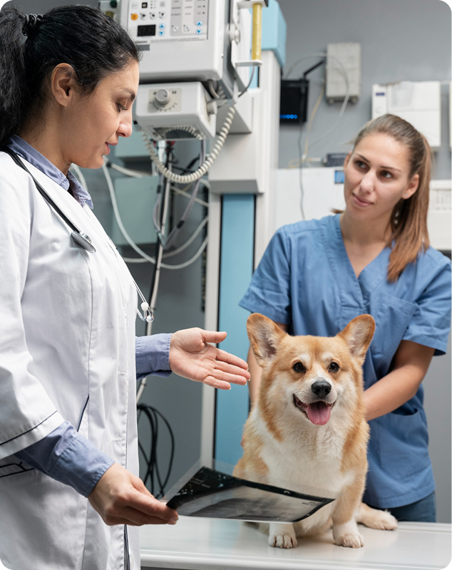
Preparing animals for MRI imaging requires accuracy, safety, and proper coordination between staff and technologists. Our clinical preparation guidelines help veterinary teams streamline workflow, reduce stress for patients, and ensure the highest quality diagnostic results. These resources provide practical steps, safety protocols, and best practices that support both new and experienced teams during every stage of the MRI process.
MRI or magnetic resonance imaging is a highly detailed field with many levels of training and safety.
This tutorial will involve aspects of MRI regarding safety measures as well as the foundation of MRI setup for patients.
Safety Precautions

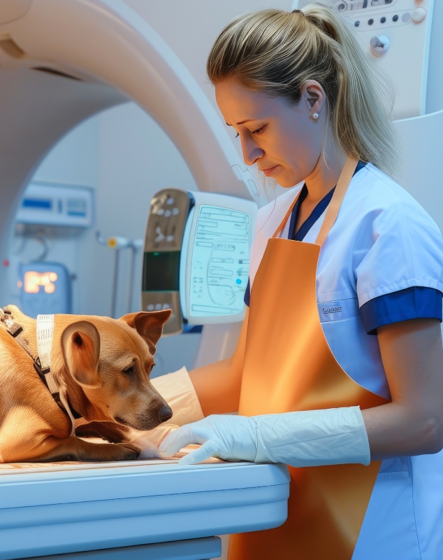
MRI or magnetic resonance imaging is a highly detailed field with many levels of training and safety. This tutorial will involve aspects of MRI regarding safety measures as well as the foundation of MRI setup for patients.
To be directly on the surface of the CTL coil, the affected leg needs to be flexed at a 90° angle where the radius ulna meets the humerus. The unaffected leg needs to be completely out of the field of view to visualize the affected leg better in the scan. The same applies with the tubing as mentioned above.

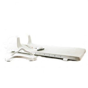
To be positioned directly around the knee of interest with the joint centered within the coil. The affected knee should be slightly flexed and comfortably supported to minimize motion during the scan. The unaffected leg must be positioned completely outside the field of view to avoid signal interference. Ensure all tubing and external accessories are routed away from the imaging area as mentioned above.
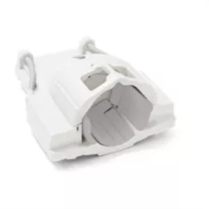
To be placed securely around the patient’s head with the head centered in the coil. The patient’s head must be positioned in a neutral alignment to reduce motion and ensure optimal image quality. Any external devices, tubing, or accessories should be kept outside the field of view. Proper padding should be used to stabilize the head throughout the scan.

To be positioned symmetrically around the anatomical area of interest with the region centered within the coil. the patient should be positioned comfortably to minimize movement during image acquisition. Any non-essential objects, tubing, or accessories must be placed outside the field of view to prevent artifacts and signal degradation.
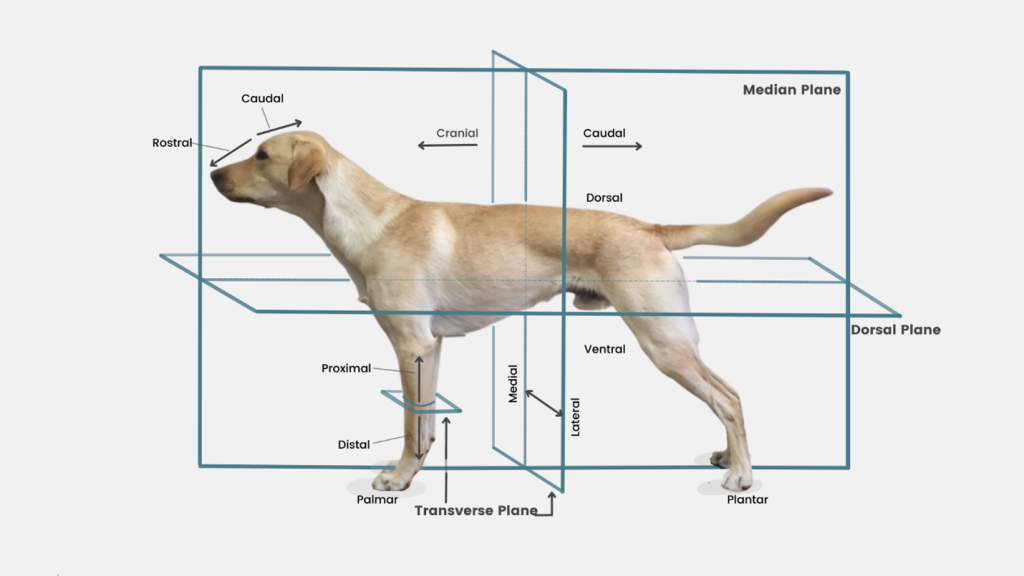
This anatomical plane reference is designed to support MRI technologists in accurately positioning patients and planning scan orientations during MRI examinations. The illustration clearly demonstrates standard anatomical planes and directional terminology as applied to veterinary anatomy, providing a consistent framework for image acquisition and interpretation. Proper understanding of these concepts is critical for producing diagnostically accurate images, maintaining protocol consistency, and ensuring effective communication between technologists and radiologists.
By using standardized anatomical references, technologists can correctly align imaging planes relative to the patient’s anatomy, reduce positioning errors, and improve reproducibility across follow-up studies. This reference is especially valuable when scanning complex regions or when adapting protocols for different patient sizes and positions.
This reference material helps MRI technologists to:
Standardized MRI protocols for small animals and exotic patients. Includes positioning, coil selection, and recommended sequences to ensure consistent, high-quality imaging.
Guidelines for veterinary contrast administration, including dosing ranges and safety considerations.
Clear positioning and coil placement guidance to reduce artifacts and improve image quality.